Overview
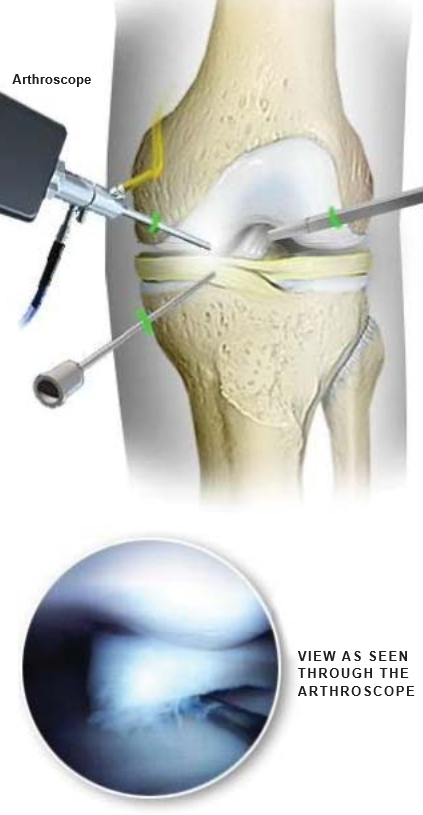
Arthroscopic surgery is used to diagnose and treat many joint problems. This significant advance in joint care allows for a rapid return to improved activity. Most commonly used in knees, shoulders and ankles, the arthroscope can also be used for the spine, hips, wrists, and elbows. This animation shows the knee joint.
Incisions Created
Three small incisions are made around the joint area. Surgical instruments will be positioned in these incisions.
Fluid Pumped into Joint
A tube-like needle is inserted in one incision. Fluid is pumped through the tube and into the joint. This expands the joint, giving the surgeon a clear view and room to work. The tube will also be used as a drainage needle to regulate the amount of fluid in the joint during the procedure.
Arthroscope Inserted
Through another incision, the surgeon inserts the arthroscope. This instrument has a light and a small video camera that sends images to a TV monitor in the operating room.
Joint Examined
With the video images from the arthroscope as a guide, the surgeon can look for damaged tissue. If the surgeon sees an opportunity to treat a problem, a variety of small surgical instruments can be inserted through the third small incision.
End of Procedure
The surgeon may close the incisions with stitches or tape. Recovery from arthroscopy is faster than recovery from traditional open joint surgery.